Thursday, July 30, 2015

40 year old female with defect #5 following extraction of cracked tooth #5


Implant Workup:

PMH: 
Healthy
Smoker: No
Diabetic: No
Periodontal Disease: No
Radiation/Bisphophonate use: No

Restorative Plan:
Single fixed: #4 and #5
fixed bridge
Implant supported overdenture
Fixed Hybrid

Hard tissue:
Vertical: 4 mm from ridge to IAN
Horizontal: defect from residual ridge down to apical third of tooth #4
Interdental space: sufficient pre-molar width 
Active infection: No
Grafting: socket preservation, gap grafting, veneer graft, guided bone regeneration

Soft Tissue: 
Biotype: Thin
Keratinized tissue: 2 mm on buccal, stable
Recession: no
Grafting: subepithelial CTgraft, free gingival graft, rotational flap, none

Smile LineHigh,  Low,  N/A

Treatment Plan
Two Stage
Single Stage
Immediate
Implant: #4: 3.5x10mm Biomet 3i bone level,  #5: 3.5x10mm Biomet 3i bone level

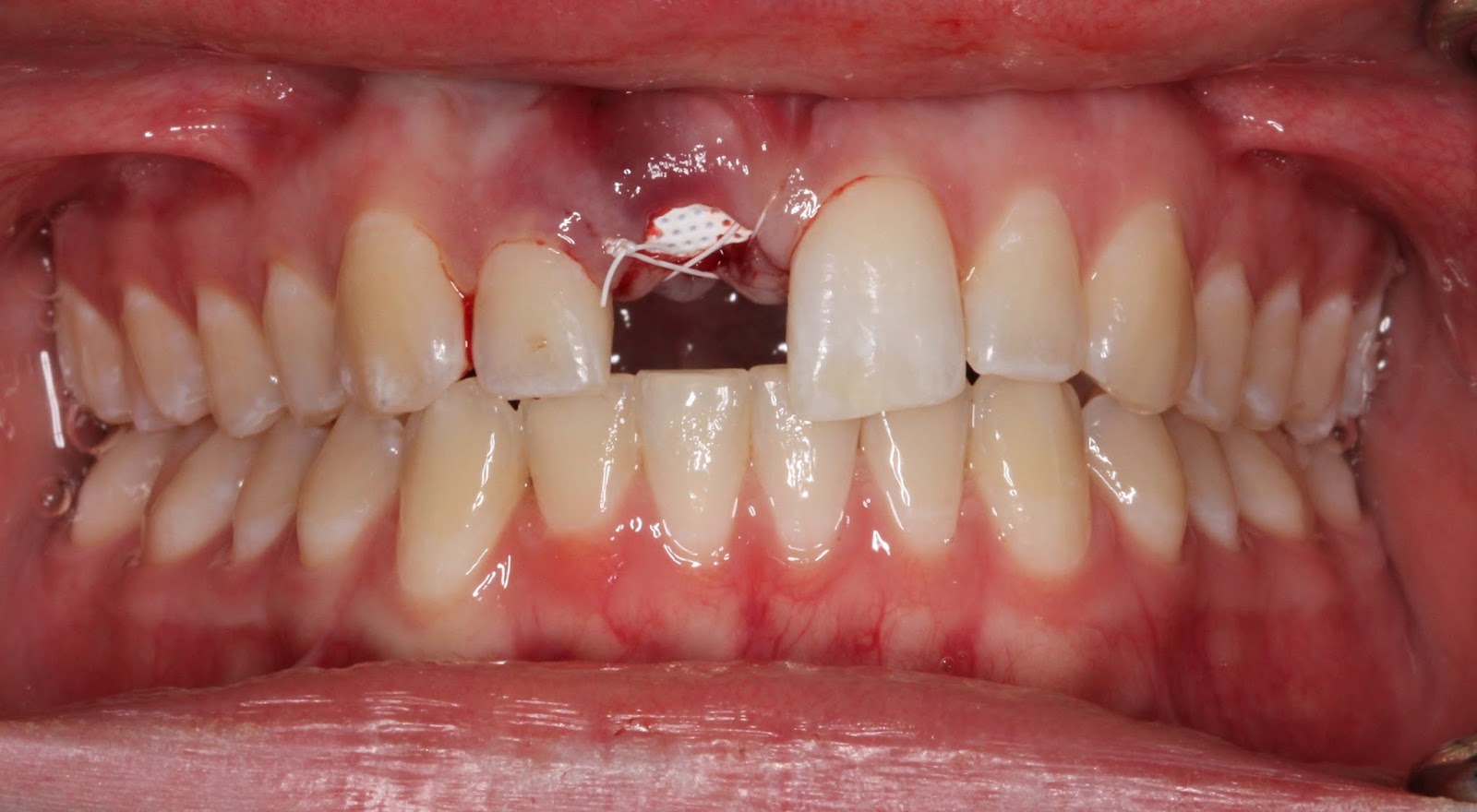
Narrative: Patient was referred for cracked tooth #5.  The tooth was extracted and socket preservation was performed, however, the graft failed and the patient was left with an unusually large hard and soft tissue defect of the site #5.  The mesial root surface of tooth #4 was exposed halfway to the apex and there was bone loss up to the apical third.  Tooth #6 still had distal bone covering most of the root surface.  Due to esthetic concerns, a bridge was not the ideal choice as it would likely need pink porcelain and would be difficult to maintain the hygiene. Because it is unpredictable to graft bone to an exposed root surface, the decision was made to extract tooth #4 and maintain the distal bone and perform guided tissue regeneration from tooth #3 to tooth #6. This was the most reasonable approach given the size of the defect and loss of bone on tooth #4.  The graft was done with MinerOss,  PRGF, and a titanium membrane. After two months healing, the membrane was removed. After an additional two months healing, the implants were placed at bone level using a CT guided stent. The implants were allowed to heal for six months and the patient is currently awaiting exposure and CT graft. The patient was given a flipper during healing of the initial extraction and an essex retainer during healing of the guided bone regeneration.  The essex gave the advantage of a buccal flange that protected the soft tissue after the membrane was removed. 

note loss of buccal convexity at site #5


note loss of mesial bone on tooth #4

Note maintenance of distal bone on the canine. 


Extraction tooth #4 and Guided Bone Regeneration site #4-5
Bone defect after full thickness flap raised

MinerOss graft mixed with PRGF

Platelet rich fibrin membrane placed over MinerOss graft

Titanium reinforced membrane secured with membrane tack

The buccal flap was coronally advanced to cover the membrane and close primarily. The membrane subsequently became exposed, but was left in place for 8 weeks.
After 2 months healing

Note the bed of granulation tissue covering the grafted area


The essix retainer was used to protect the formation of new gingiva.


After 4 months healing

Note the increased height and width of the grafted site. 


Note the formation of new keratinized gingiva over graft site and the augmented buccal width

Note the gain in vertical height.

Implant Placement #4, #5.


A CT guided stent was used for the pilot drill.

BioMet 3i tapered bone level implants both 3.5x10mm

The gingival was sutured over the implants and will be allowed to integrate for four months.  At the time of uncovery a connective tissue graft will likely be necessary to attempt to recreate and mold a papilla between the new prosthesis and the existing teeth. 


Wednesday, July 29, 2015

25 year old female with internal root resorption tooth #8


Implant Workup:

PMH: 
Healthy
Smoker: No
Diabetic: No
Periodontal Disease: No
Radiation/Bisphophonate use: No

Restorative Plan:
Single fixed
fixed bridge
Implant supported overdenture
Fixed Hybrid

Hard tissue:
Vertical: 15 mm from ridge to nasal floor
Horizontal: 5 mm ridge width
Interdental space: sufficient incisor width 
Active infection: Yes
Grafting: socket preservation, gap grafting, veneer graft, guided bone regeneration

Soft Tissue: 
Biotype: Thin
Keratinized tissue: 3 mm on buccal, stable
Recession: no
Grafting: subepithelial CTgraft, free gingival graft, rotational flap, none

Smile Line: High,  Low,  N/A

Treatment Plan
Two Stage
Single Stage
Immediate
Implant: #9 BioMet tapered platform switch 3.5 x13mm 

Narrative:
This young women was referred for extraction and implant tooth #8. The tooth had internal resorption and had a root canal over a year previously.  The resorption continued until the tooth was deemed non-restorable and had a buccal parulus.  The decision was made to extract the tooth and perform socket preservation due to the active infection.  The patient was previously on antibiotics and had no purulence on the day of surgery. After 4 months healing a CT scan was taken which showed the residual ridge had only 5 mm of facial-palatal width.  She also had a high smile line and very thin gingival biotype.  On the day of surgery, a mid crestal incision was made and a split thickness flap was developed on the facial for a sub-epithelial connective tissue graft. The palatal flap was designed sub-periosteally allowing for placement of the implant.  The bone level implant was placed in the cingulum region of tooth #8 and the CT graft, harvested from the tuberosity was placed in the facial recipient site and sutured with 4.0 chromic gut. In the pictures, the bulk that was achieved from the CT graft is evident.

Pre-operative Photos
Note the healing parulus on the distobuccal root #8

Note the high smile line showing the anterior papilla and entire crowns of the laterals

Note the resorption on the palate of tooth #8


MinerOss and Cytoplast membrane for socket preservation with 4.0 PTFE sutures

Flipper in place
4 Months Healing

Note mild granulation tissue -- likely from flipper use/trauma

Note the loss of facial convexity site #8

Implant #8 with sub-epithelial CT graft
Implant placed at cingulum #8--bone level--buried II stage approach. 

CT graft harvested from Tuberosity.  All epithelium removed. 

Instead of using a papillary sparing incision, a crestal incision was made to accomodate a split-thickness flap and placement of a CT graft.  This could also be done at the uncovery appointment. 

Note the facial convexity obtained by the CT graft. 
Post Op Panorex
Implant is placed 3 mm apical to the free gingival margin. 


Friday, July 24, 2015

40 year old male immediate implant #30


Implant Workup:

PMH: 
Healthy
Smoker: No
Diabetic: No
Periodontal Disease: No
Radiation/Bisphophonate use: No

Restorative Plan:
Single fixed
fixed bridge
Implant supported overdenture
Fixed Hybrid

Hard tissue:
Vertical: 14 mm from ridge to IAN
Horizontal: 8 mm ridge width
Interdental space: sufficient molar width 
Active infection: No
Grafting: socket preservation, gap grafting, veneer graft, guided bone regeneration

Soft Tissue: 
Biotype: Thick
Keratinized tissue: 3 mm on buccal, stable
Recession: no
Grafting: subepithelial CTgraft, free gingival graft, rotational flap, none

Smile Line: High,  Low,  N/A

Treatment Plan
Two Stage
Single Stage
Immediate
Implant: #30 BioMet 5.0x 11.5mm standard platform

Narrative: Because of the short retained roots and absence of active periodical infection, it makes sense to do an immediate implant at this site.  There is little benefit to grafting when remaining bone after extraction is sufficient to provide primary stability. The remaining gap on the mesial and distal of the implant was grafted with BioOss cancellous particles covered by a  collagen membrane and closed with 4.0 silk.