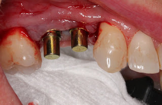
Implant Workup:
PMH:
Healthy
Smoker: No
Diabetic: No
Periodontal Disease: No
Radiation/Bisphophonate use: No
Restorative Plan:
Single fixed: #4 and #5
fixed bridge
Implant supported overdenture
Fixed Hybrid
Hard tissue:
Vertical: 4 mm from ridge to IAN
Horizontal: defect from residual ridge down to apical third of tooth #4
Interdental space: sufficient pre-molar width
Active infection: No
Grafting: socket preservation, gap grafting, veneer graft, guided bone regeneration
Soft Tissue:
Biotype: Thin
Keratinized tissue: 2 mm on buccal, stable
Recession: no
Grafting: subepithelial CTgraft, free gingival graft, rotational flap, none
Smile Line: High, Low, N/A
Treatment Plan
Two Stage
Single Stage
Immediate
Implant: #4: 3.5x10mm Biomet 3i bone level, #5: 3.5x10mm Biomet 3i bone level
Narrative: Patient was referred for cracked tooth #5. The tooth was extracted and socket preservation was performed, however, the graft failed and the patient was left with an unusually large hard and soft tissue defect of the site #5. The mesial root surface of tooth #4 was exposed halfway to the apex and there was bone loss up to the apical third. Tooth #6 still had distal bone covering most of the root surface. Due to esthetic concerns, a bridge was not the ideal choice as it would likely need pink porcelain and would be difficult to maintain the hygiene. Because it is unpredictable to graft bone to an exposed root surface, the decision was made to extract tooth #4 and maintain the distal bone and perform guided tissue regeneration from tooth #3 to tooth #6. This was the most reasonable approach given the size of the defect and loss of bone on tooth #4. The graft was done with MinerOss, PRGF, and a titanium membrane. After two months healing, the membrane was removed. After an additional two months healing, the implants were placed at bone level using a CT guided stent. The implants were allowed to heal for six months and the patient is currently awaiting exposure and CT graft. The patient was given a flipper during healing of the initial extraction and an essex retainer during healing of the guided bone regeneration. The essex gave the advantage of a buccal flange that protected the soft tissue after the membrane was removed.
 |
| note loss of buccal convexity at site #5 |
 |
| note loss of mesial bone on tooth #4 |
 |
| Note maintenance of distal bone on the canine. |
Extraction tooth #4 and Guided Bone Regeneration site #4-5
 |
| Bone defect after full thickness flap raised |
 |
| MinerOss graft mixed with PRGF |
 |
| Platelet rich fibrin membrane placed over MinerOss graft |
 |
| Titanium reinforced membrane secured with membrane tack |
 |
| The buccal flap was coronally advanced to cover the membrane and close primarily. The membrane subsequently became exposed, but was left in place for 8 weeks. |
After 2 months healing
 |
| Note the bed of granulation tissue covering the grafted area |
 |
| The essix retainer was used to protect the formation of new gingiva. |
After 4 months healing
 |
| Note the increased height and width of the grafted site. |
 |
| Note the formation of new keratinized gingiva over graft site and the augmented buccal width |
 |
| Note the gain in vertical height. |
 |
| A CT guided stent was used for the pilot drill. |
 |
| BioMet 3i tapered bone level implants both 3.5x10mm |


No comments:
Post a Comment